
Gallery 25: Infection of H-9 cells with the MN strain of HIV-1 virus, electromicrograph 3
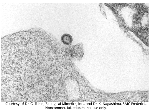
Virus particle budding out from the cell. Although similar to the previous micrograph, the inner core is not as dense and therefore this is an "immature" viron budding out as opposed to a mature virus fusing in to infect a cell.
virus particle, viron, micrograph, inner core, hiv, cells
- ID: 16560
- Source: DNALC.DNAFTB
Related Content

16559. Gallery 25: Infection of H-9 cells with the MN strain of HIV-1 virus, electromicrograph 2
Virus particle is fusing with the cell membrane and about to empty its contents into the cell. Note the visible inner core.

16558. Gallery 25: Infection of H-9 cells with the MN strain of HIV-1 virus, electromicrograph 1
Magnification including computer enhancements are approximately 100,000 times. A mature round virus particle is sitting next to the cell ready for infection. Note the visible inner core.

16561. Gallery 25: Infection of H-9 cells with the MN strain of HIV-1 virus, electromicrograph 4
Mature virus particles released from host cell.

16135. HIV jumps barrier zoonosis
Image showing Watson and Crick, chimp, macaques, HIV virus particles.

16137. HIV Virion annotated
HIV particle proteins annotated.

16552. Animation 25: Some viruses store genetic information in RNA.
David Baltimore and Howard Temin explain work on the Rous sarcoma virus.

16085. HIV/SIV Genome Maps
Contains an image depicting the genome maps of HIV-1 HXB2, HIV-2 BEN, and SIV Sykes.

16138. HIV artistic rendering
HIV artistic rendering.

16649. Gallery 30: An electron micrograph of a mouse liver cell
An electron micrograph of a mouse liver cell. Magnification approximately 12,000 times.

16231. Gallery 7: Micrograph of Cell Dividing, 2
(2 of 4) Cell dividing: chromosomes are visible and lined up at the plane of division.