
Micrograph of Cell Dividing, 2
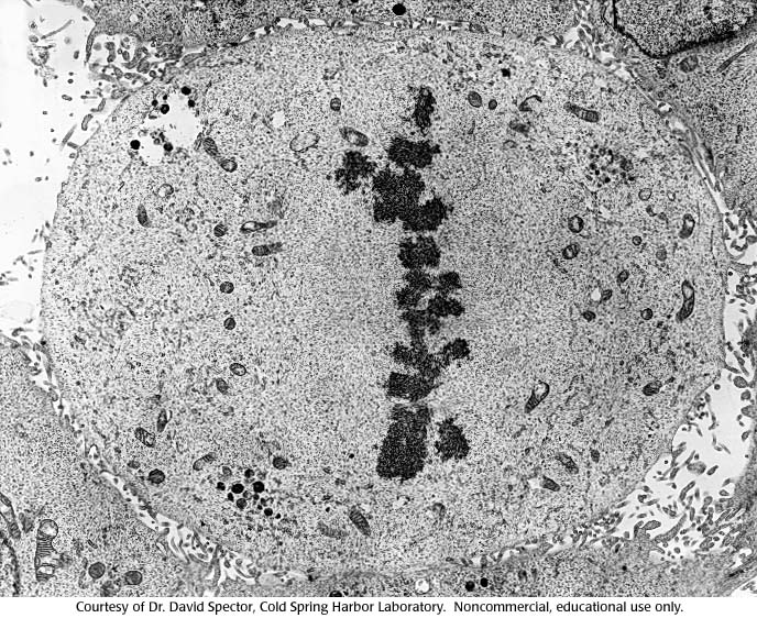
(2 of 4) Cell dividing: chromosomes are visible and lined up at the plane of division.
micrograph, chromosomes, gallery 7
- ID: 16231
- Source: DNALC.DNAFTB
Related Content

16232. Gallery 7: Micrograph of Cell Dividing, 3
(3 of 4) Cell dividing: chromosomes are being pulled toward the cellular poles.

16638. Gallery 29: Chromosome with histone stripped
Electron micrograph of the DNA and the protein scaffold left over from one chromosome (insert) with all the histone stripped out.

16636. Gallery 29: Electron micrograph of chromatin
Electron micrograph of the 10-nm fiber.

16230. Gallery 7: Micrograph of Cell Dividing, 1
(1 of 4) Photomicrograph of a cell dividing: nucleus is visible as dark staining organelle.

16637. Gallery 29: Electron micrograph of chromatin (1)
Electron micrograph of the 30-nm fiber.

16767. Gallery 37: Normal Drosophila Head, electron micrograph
Scanning electron micrograph of the head a normal Drosophila.

16233. Gallery 7: Micrograph of Cell Dividing, 4
(4 of 4) Cell dividing: mother cell divides resulting in two daughter cells each with its own nucleus.

16649. Gallery 30: An electron micrograph of a mouse liver cell
An electron micrograph of a mouse liver cell. Magnification approximately 12,000 times.

16768. Gallery 37: Antennapedia Drosophila Head, electron micrograph
Scanning electron micrograph of the head a Drosophila mutant for the antennapedia gene.

16533. Gallery 24: Electron micrograph of RNA/DNA hybrid
This was one of the original photos that Roberts and his group used for analyzing their results.